Case Study: Luxating Patella Surgery
21st May 2021 | Posted by The Team at Coquet Vets
Bella all cosy in her kennel before surgery. (Image: Coquet Vets, 2021)
Meet Bella, a 10-month-old Cavalier cross. Her owners noticed that when she was walking, she was holding up her left hind leg for a few seconds, and then placing back down. This had been going on for a few days.
Our vet, Emily, checked Bella over and she could feel the patella (kneecap) in Bella’s left leg popping in and out. This is known as a luxating patella. This was also the same with Bella’s right leg. Emily advised strict rest and pain relief to go home with, and advised to book in for X-rays so we could confirm the diagnosis.
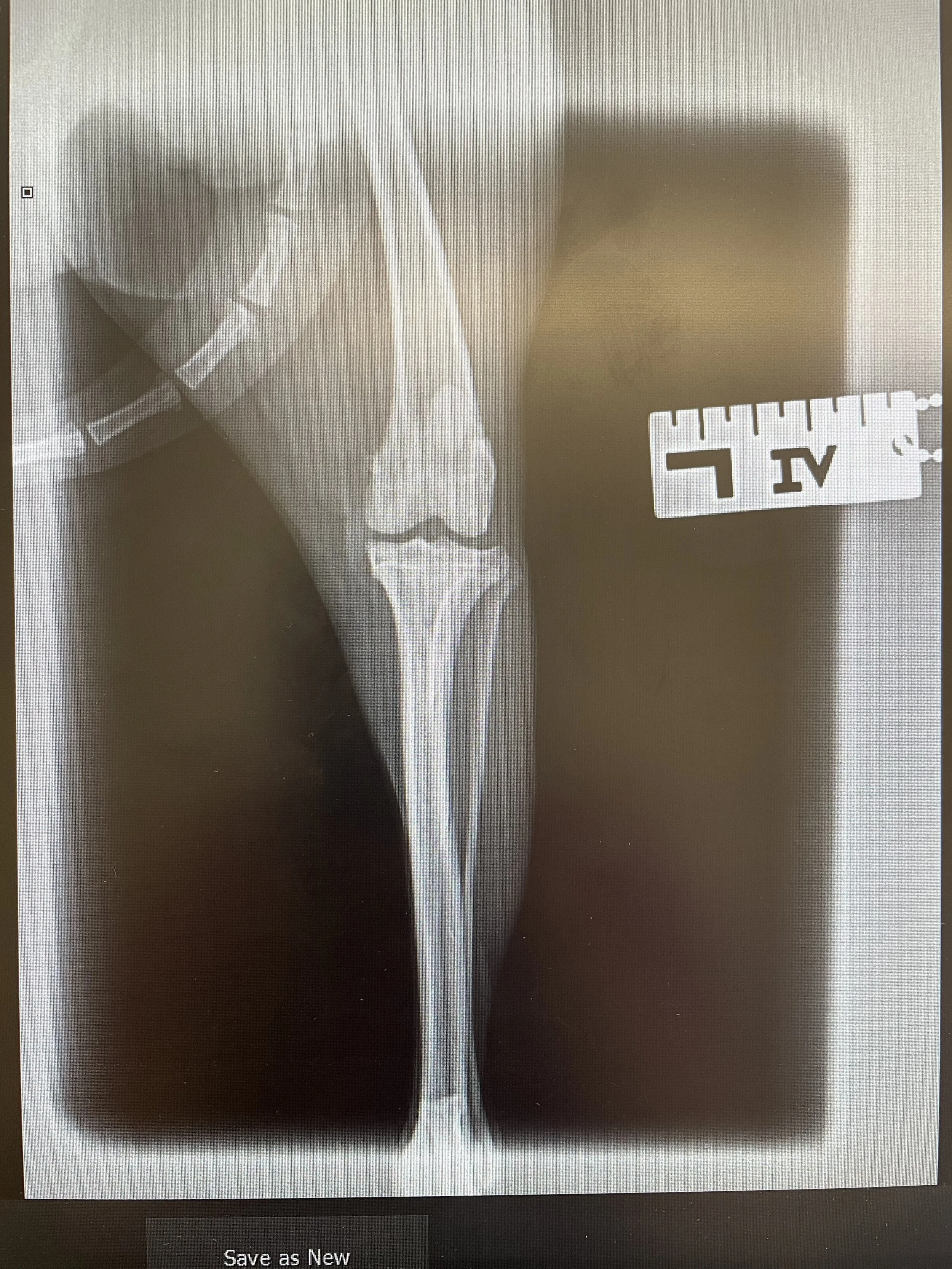
A few days later Bella came in for a general anaesthetic and X-rays of both of her hind legs and hips. An X-ray is a quick and painless procedure commonly used to produce images of the inside of the body. It's a very effective way of looking at the bones and can be used to help detect a range of conditions.
This is the before surgery X-ray (Image: Coquet Vets, 2021)
Multiple X-rays were taken. We took two views of each leg, a lateral position, which is the patient lying on the right or left side, as well as a view called ‘dorso-ventral’, which is the patient lying on their front and we stretch the leg out backwards. The X-ray head is positioned over the area that we want to X-ray (in this case the stifle and hips).
We sent all of the X-rays to Neil Adams, our visiting orthopaedic surgeon. He has been doing orthopaedic surgeries for fifteen years. Neil advised that Bella needed surgery, and he recommended operating on the worst leg (left) first, but that it was likely she will need to have both legs operated on. Luxating patella’s don’t always need surgical treatment, but due to the frequency of Bella’s luxation, surgery was indicated.
Neil in the operating theatre with Bella. (Image: Coquet Vets, 2021)
After speaking to Neil, a date was confirmed to go ahead with patella surgery on the left leg. Neil performed the surgery by deepening the trochlear groove (the patella sits in this) and performed a tibial tuberosity transposition. This is corrective surgery to secure the patella into the trochlear groove. This was stabilised with wires and then the wound was sutured closed. We always take post-operative X-rays to confirm the wire is in the correct place.
Sterile area and leg prepped (Image: Coquet Vets, 2021)
Post-operation X-ray (Image: Coquet Vets, 2021)
After the X-rays were checked, Bella was woken up and put into a kennel to recover. Her recovery was uneventful, and she went home later that day.
Bella went home with pain relief and an exercise plan for the next 12 weeks. Bella was also scheduled for a post-operative check with a vet 5 to 7 days later to see how the leg is healing after surgery. After 8 weeks, Bella will come back in for X-rays to check everything is healing correctly.
If this article has raised any questions or concerns, contact us on 01665 252 250 or email us at info@coquetvets.co.uk.


